Sections

“There’s something wrong with her head.” 

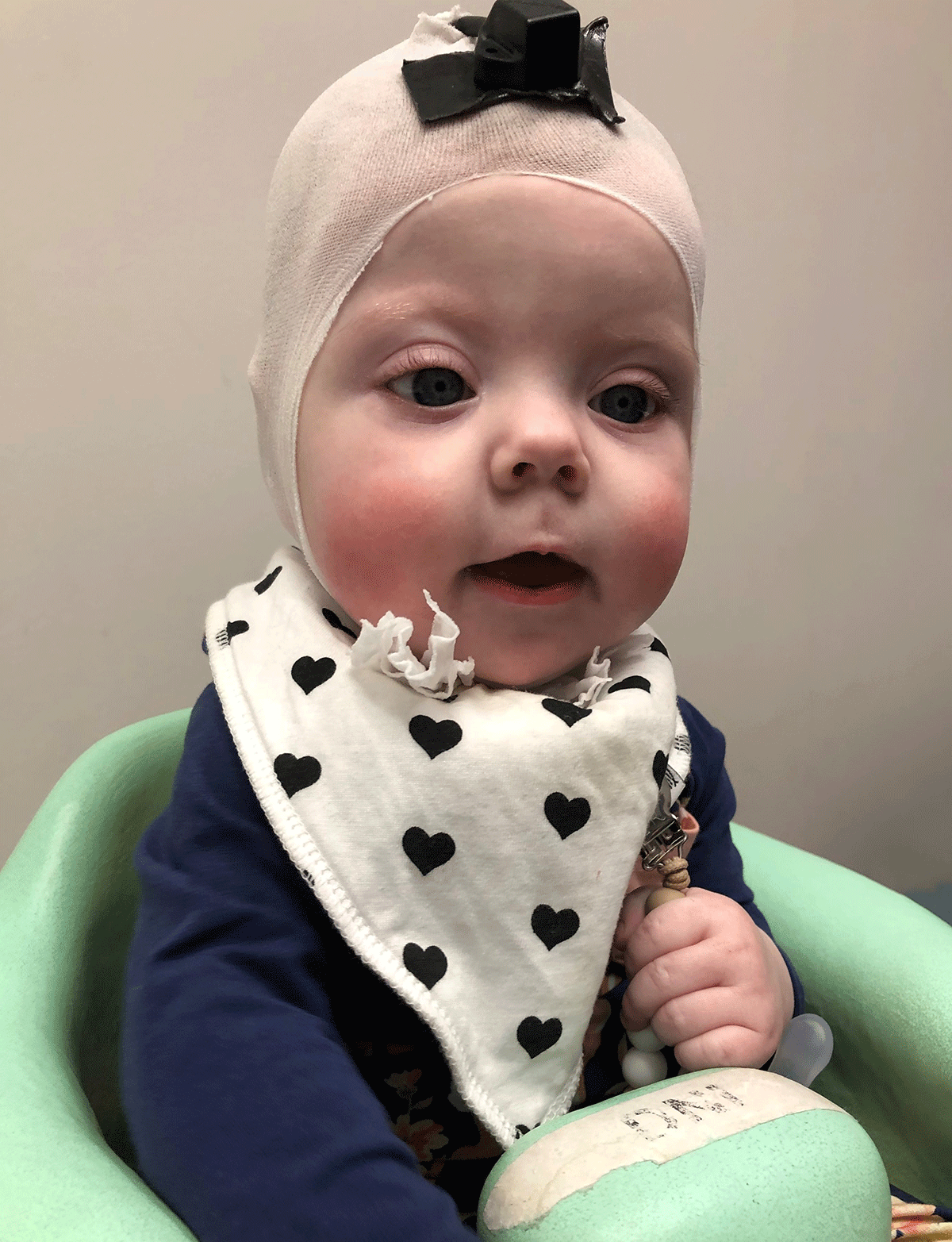
Megan Kane was not prepared for those words from her husband, Mark. Looking closer at their newborn, Imogene, the North Liberty, Iowa, couple noticed that the right side of her forehead didn’t appear to be as full as the left. 

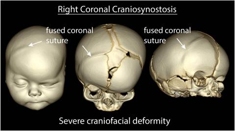
Two weeks later, Imogene’s pediatrician ordered an X-ray that confirmed their fear: Their baby girl had a fusion of the right coronal suture—two plates in her skull had fused together prematurely and would need to be separated.

Imogene, the Kanes' second child, had been born with craniosynostosis, a condition that traditionally would have meant hours of surgery. But because Imogene was diagnosed early after birth and seen quickly at the University of Iowa Stead Family Children’s Hospital, Brian Dlouhy, MD (13R, 14F), pediatric neurosurgeon, and Deborah Kacmarynski, MD, pediatric otolaryngologist, were able to employ a relatively new, minimally invasive procedure that significantly reduces the length of surgery and minimizes blood loss.

Image
Preoperative 3d CT

New option results in shorter surgery time

Craniosynostosis is a birth defect in which one or more of the seven fibrous joints in a baby’s skull—called sutures—is prematurely closed. This limits the space needed for brain and skull growth and often leads to craniofacial malformations. 

The condition affects approximately 1 in 2,000 babies each year, and is likely caused by a combination of environmental, hormonal, and genetic factors that make the skull suture a little more likely to fuse, according to Saul Wilson, MD (09R, 10F), a pediatric neurosurgeon at UI Stead Family Children’s Hospital.

Surgeons at the children’s hospital see about 35 to 40 cases of craniosynostosis of varying degrees each year, says UI pediatric neurosurgeon Arnold Menezes, MBBS (74R). 

Traditionally, the condition is repaired with an open surgery, in which a team of surgeons from pediatric neurosurgery, otolaryngology, and plastic and reconstructive surgery make an incision from ear to ear across the top of the skull and remove sections of the skull that have fused together, preferably within the first three to six months after birth, Menezes says. Repairs are often made to any craniofacial abnormalities at the same time. The surgery can take several hours and typically means a three- to five-day stay at the hospital.

These kids with the endoscopic procedure are typically leaving the hospital the day after surgery.”
Brian Dlouhy, MD, assistant professor of neurosurgery

Recently, however, that same surgery team at Iowa began offering an endoscopic procedure for infants with craniosynostosis. Dlouhy, who was trained on the newer technique, says the procedure can be completed in less than an hour. Rather than requiring a full incision on the scalp to create large bone cuts, which result in significant tissue and bone manipulation, a smaller incision is made near the fused suture. A small camera is used to guide the surgeon in removing the bone.

While the open surgery is usually done when the baby is between 3 and 6 months old, the endoscopic procedure is performed before the infant is 3 months old. Being able to address the issue early allows the brain and skull more time to grow together naturally, according to Dlouhy.

“These kids with the endoscopic procedure are typically leaving the hospital the day after surgery,” he says. “Swelling is very minimal, they need less anesthesia, and it's less traumatic for the baby.”

Image
8 weeks old, 1 day before surgery
Image
Imogene Kane, 4 days after surgery
4 days after surgery
Image
6 months old, fitting for 3rd helmet
6 months old, fitting for third helmet

Parents play an important role in recovery

Dlouhy notes, however, that the minimally invasive approach places more responsibility on the parents. With the open surgery, a team of surgeons works to shape the skull and facial features, whereas the endoscopic procedure addresses only the fused suture. As a result, the infant is required to wear a helmet for up to a year to help the skull form properly.

Because a baby’s head grows so much during their first year—the brain typically triples in size during this period—the helmet must be resized often, sometimes weekly.

“It’s a commitment, it really is,” Megan Kane says. “You have to get it refitted every two weeks.”

By the time she was 6 months old, Imogene was on her third helmet. Kane says Imogene has adapted well to the helmet, which she’s required to wear 23 hours a day. The Kanes had a local artist paint it with flowers to look more like an accessory.

“She's been doing really well since right after the procedure,” Kane says of her daughter. “There was no blood loss, and she was able to breastfeed right away that night. Everything has been fine.”

“The molding helmet requires that the parents really be religious about their follow-through,” says UI plastic and reconstructive surgeon Mark Fisher, MD. 

Nonetheless, Dlouhy says that for patients who qualify, the endoscopic procedure is an effective alternative to the full open surgery—good news for parents and their babies. 

Image
Gold medal

Pediatric Neurosurgery

University of Iowa Stead Family Children’s Hospital is home to one of the most experienced and comprehensive pediatric neurosurgery programs in the nation and the only nationally ranked program in Iowa.