

Learn more about the four different approaches
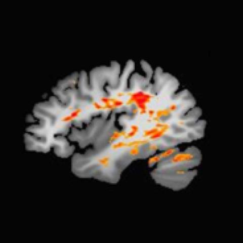
Brain imaging
Conducting brain imaging on the same research participants over time and at different stages of their illness is helping Iowa researchers better understand the nuances of bipolar disorder.
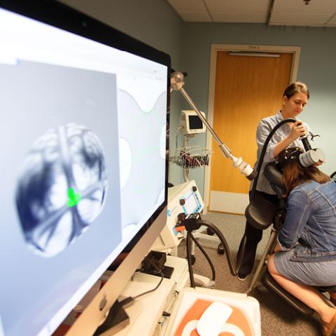
Cerebellum-targeted TMS
Researchers are still in the discovery phase of using repetitive transcranial magnetic stimulation (rTMS) directly on the cerebellum as a novel treatment option for bipolar disorder.

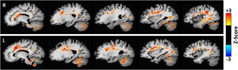
Modeling a bipolar brain
Using electrophysiology, researchers are measuring brain activity and connectivity—in hopes of better understanding exactly how the cerebellum works, and to possibly reveal its role in bipolar disorder.
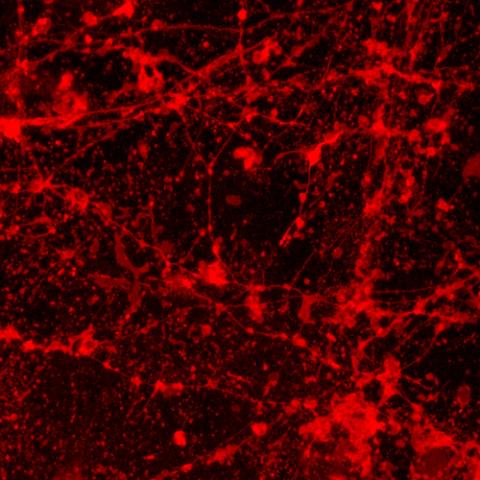
Stem cells and genetic approaches
An Iowa team is using patients’ skin cells to create stem cells, which are then turned into neurons. These neurons might offer insights into...
Brain imaging
Cerebellum-targeted TMS
Modeling a bipolar brain

Uncovering brain disorders
The Iowa Neuroscience Institute is fostering collaborations among researchers to find causes of— and the preventions, treatments, and cures for—the many diseases that affect the brain and nervous system.
Stem cells and genetic approaches
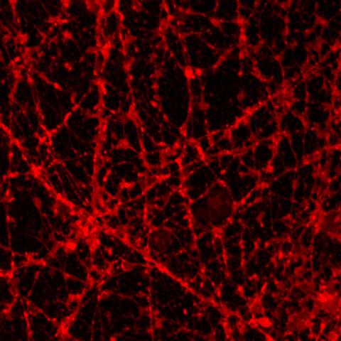
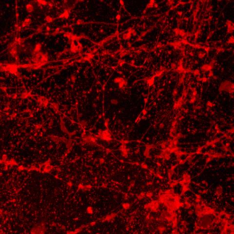
speckled appearance—compared to the control image. Neuronal beading is typically seen as a result of mitochondrial dysfunction which has previously been hypothesized in bipolar disorder. Images submitted by Karina Kruth, PhD, postdoctoral fellow.