Sections

Our brains control almost every aspect of our lives but exactly how it’s done—especially when it comes to functions like speech—is still largely a mystery.  

Researchers at the University of Iowa are working to change that.  

By building on nearly 30 years of groundbreaking research about the human brain, Iowa’s Human Brain Research Lab incorporates new and innovative techniques to study brain physiology. The lab's discoveries are paving new avenues to how researchers study brain structure and function.  

Iowa researchers are particularly interested in what happens in the brain when things function abnormally.

“If we understand how it’s supposed to work normally, it gives us a chance to correct things when they’re not normal,” says Jeremy Greenlee, MD (04R, 05F), professor in the UI Department of Neurosurgery.

The outcomes of unlocking these mysteries could be life-changing for millions of people.  

“If we come up with some kind of way to restore abnormal brain circuits and improve function, that would be the holy grail outcome of this research,” says Greenlee, who serves as the Arnold H. Menezes Chair in Neurosurgery. 

Image
electrodes in the brain

The images show the proportion of electrodes displaying speech planning responses. Most (95%) planning-related electrodes were grouped in a frontotemporal region, with the highest densities of planning electrodes clustered in the caudal inferior frontal gyrus (cIFG), or Broca’s region, and the caudal middle frontal gyrus (cMFG). A smaller number of planning sites were located in the ventral speech motor cortex (vSMC) and the anterior superior temporal gyrus (aSTG).

Those insights could lead to new treatments that would help patients with a range of conditions—including epilepsy, Parkinson’s disease, stuttering—as well as those recovering from a brain injury, stroke, or brain tumor removal, which can impact the way an individual processes information and communicates.  

This research requires going beyond imaging the brain. Investigators are using experimental methods that directly interact with the brain, including intracranial electrocorticography, deep brain stimulation, and brain cooling. They typically work with neurology and neurosurgery patients who volunteer to be part of their research during procedures they are already scheduled to undertake.  

“When you’re trying to understand how the human brain works, there are noninvasive research methods like MRI and scalp EEG that are very important, but there are certain key scientific questions that you can’t answer unless you have devices that are in direct contact with the brain,” says Matthew Howard, MD, professor and chair of the Department of Neurosurgery.  

Image
Jeremy Greenlee, MD
Jeremy Greenlee, MD

In a 2022 Nature paper, Iowa researchers applied these tactics to gain a better understanding of the brain networks that are responsible for the snappy repartee of natural conversation.

Researchers played word games with patients who were awake while their neural activity was monitored with electrocorticography electrodes placed on the surface of the brain.  

They identified two areas of the brain (Broca’s region and the caudal middle frontal gyrus, or cMFG) that are associated with speech planning, or thinking about what to say. Although previous research had shown that the Broca’s region was important for language, cMFG had not previously been linked to speech planning. 

The discovery of this circuitry could help researchers better understand communication disorders like stuttering and apraxia, where speech planning is disrupted or abnormal.  

“How we produce speech and correct for errors in real time are poorly understood but important for social beings,” Greenlee says. “If we understand how we do that, we could help people who have lost that ability.”  

The findings are also applicable for patients with neurodegenerative disorders and those recovering from brain injury or stroke.  


Iowa build major collaborations to better understand speech, improve research methods

The Human Brain Research Lab has also perfected a study technique called brain cooling, in which miniature devices are placed on parts of the brain to cool spots roughly the size of a quarter. It works much like electrical stimulation, where it temporarily turns off that part of the brain. Iowa is a leader in this kind of technical capability, which has resulted in partnerships with researchers around the world, including a decade-long collaboration with researchers at New York University School of Medicine. 

In a 2020 paper published in World Neurosurgery that resulted from this collaboration, researchers showed that brain cooling can be used to identify and protect areas of the brain during neurosurgery. In a trial with patients who were already undergoing brain surgery at UI Hospitals & Clinics, they cooled parts of the brain associated with speech. The research team found that speech was slow and slurred while these specific brain regions were cooled. But after being warmed back up, speech returned to normal, indicating the brain was unaffected by the process. That information helped the neurosurgeons protect the speech centers during surgery.   

This discovery has opened a new avenue of studying the brain without the risk of seizure that electrical cortical stimulation (ECS), a more common technique, does. It proved the safety of the technique, and that it can continue to be used in the kind of research the Human Brain Research Lab is known for.   

"We showed that brain cooling is a safe alternative to ECS, and it allows us to be a little bit more focused with what areas of the brain we’re affecting", Greenlee says.  "We’ve been working with colleagues from New York University for more than 10 years, and it’s been fun to see the evolution of that research."

Our patients are not going to benefit from the data we get, or things we’re looking at from them, but they work with us because they hope that it translates into helping people like them down the road, Greenlee says.”
Jeremy Greenlee, MD

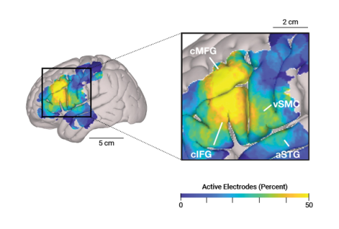
While this work is rooted in neurosurgery, it crosses disciplines at Iowa, Greenlee says.  

“We do a lot of research with Parkinson’s disease patients in trying to improve speech outcomes, which is a collaborative effort with people in psychology and neurology,” he says. 

In 2021, a collaborative team of researchers at Iowa received a $3.1 million grant from the National Institutes of Health (NIH) to study changes in speech and look at movement in Parkinson’s patients who undergo bilateral deep brain stimulation. 

And this isn’t the first time the NIH has funded Iowa’s brain physiology research. The UI was awarded a Human Auditory Cortex Physiology grant in 1995 that has been competitively renewed through 2025.    

“It may be the longest continuously funded R01 grant by a neurosurgeon principal investigator in the U.S.,” says Howard, who was an assistant professor when the grant was initially awarded. 

Greenlee and Howard recognize that they wouldn’t be able to do this kind of research without the generosity of their neurosurgery patients. 

“Our patients are not going to benefit from the data we get, or things we’re looking at from them, but they work with us because they hope that it translates into helping people like them down the road,” Greenlee says.