Amyloids can be found in many neurodegenerative diseases, and several proteins can form amyloids—including tau, beta amyloid, and alpha synuclein. The left image shows that the tangles and neuritic plaques in Alzheimer’s disease are amyloids because the magenta and green staining overlap. New PET imaging tracers being developed to diagnose neurodegenerative diseases detect these amyloids using dyes that are derived from thioflavin.
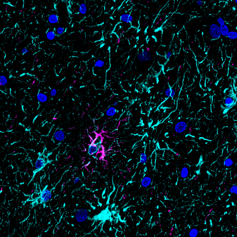
The right image shows the brain cortex of a patient with progressive supranuclear palsy (PSP), a disease that affects the frontal and temporal lobes of the brain and is part of a larger group of diseases called frontotemporal dementias. PSP is another disease that is diagnosed by the accumulation of tau protein (magenta), which is found in cells called astrocytes (cyan). Unlike what’s seen in Alzheimer’s disease, there’s no thioflavin (green) staining in the astrocytic or neuronal tau accumulations, suggesting that the protein aggregates in PSP are not amyloids.
These images are included in a 2023 paper published in the Journal of Histochemistry and Cytochemistry showing that across all frontotemporal dementias, none have thioflavin-positive amyloids (green) despite tau staining. Why amyloids form in certain diseases, such as Alzheimer’s, and not in others, such as PSP, is not well understood, but this research suggests that new imaging tracers based on thioflavin S derivates won’t work for certain diseases.