Stead Family Scholars program bolsters early-career faculty
New research funding program supports young investigators, pushes "risky" research
Five investigators were named to the inaugural cohort of the Stead Family Scholars program, which aims to recognize and advance the development of outstanding early-career faculty.
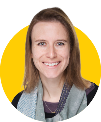
“It really shows that the College of Medicine is supporting what we do,” says Lilliana Radoshevich, PhD, assistant professor of microbiology and immunology. “The Stead family has supported groundbreaking new research that can generate data that is applicable for National Institutes of Health (NIH) grants. The importance of that can’t be underscored enough.”
The program will give a boost to help “risky” projects into the next phase of research.
“The funding from the Stead Family Scholars allows us to address questions that are hugely important, but that might not be fundable through the NIH for a couple more years,” says Mary Weber, PhD, MS, assistant professor of microbiology and immunology. “It will allow us to fill in those gaps to build a strong case for outside funding.”
Each investigator will receive $125,000 in annual research funding for three years.
“Science is about discovery, but you’re often limited in what you can do financially,” says Ling Yang, PhD, assistant professor of anatomy and cell biology. “This funding gives us the resources, the foundation, and the passion to really explore.”
The scholars also receive professional development training. The group was selected for their contributions to the college’s tradition of research excellence.
“I’m proud to be able to represent translational medicine in the group of scholars,” says Jennifer Bermick, MD, associate professor of pediatrics. “It’s a difficult field, and a difficult field to get recognition for.”
By fostering these nascent discoveries, the Stead family and the Carver College of Medicine is investing in the future of patient care.
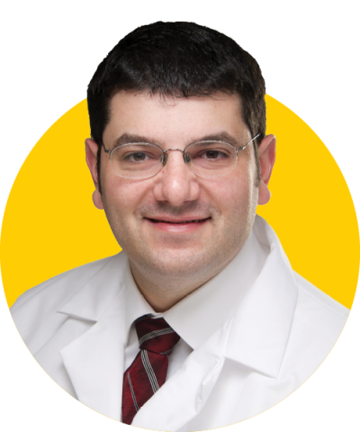
“I’m excited about the ability to accelerate the advances we’re making,” says Munir Tanas, MD, associate professor of pathology. “Hopefully having this support will allow us to move more quickly toward helping patients.”

Jennifer Bermick, MD
Associate Professor of Pediatrics–Neonatology
Many neonatal therapies are less effective because we still don’t fully understand babies’ immune systems. Bermick’s lab is expanding our understanding so we can develop more targeted therapies for neonates in the future.
Her efforts are focused on infection and sepsis, which account for 1 million infant mortalities worldwide per year.
“At Iowa, we’re fortunate to have the best extremely pre-term baby outcomes in the country,” Bermick says. “These babies do well, and I can follow them longitudinally to do immune profiling and figure out what normal levels of inflammation are and how early-life exposure to infection or antibiotics influences them.”
She has observed that 34 weeks may be an important milestone in immune development—a finding Bermick says no one has studied before.
“If we can figure out what’s regulating that change, we can push them a little bit sooner,” she says.
Milestone publications

Lilliana Radoshevich, PhD
Assistant Professor of Microbiology and Immunology
Radoshevich studies the properties of ubiquitin-like proteins to understand how cells defend against infection. The protein ISG15 is her main target of study.
“No one knows what it does, but it’s dysregulated in cancer, neurodegenerative disease, and autoimmune disease,” Radoshevich says. “If we can learn what it’s doing in the context of bacterial infection, we can see if it would be a good target for other difficult-to-treat human diseases.”
To do so, her research team is identifying the functions of a family of intracellular protein “tags” that modify other proteins.
“We think if we can learn to activate one of them, it could potentially make cells migrate less well. This could affect metastasis in the cancer setting,” Radoshevich says.
Someday, this could translate to immune-boosting drugs that could protect against hypothetical viral epidemics in the future. In the meantime, the team is building the understanding of the pathways that viral pathogens can hijack, which has broad implications for managing disease and developing therapies.
Milestone publications
-
Ring finger protein 213 assembles into a sensor for ISGylated proteins with antimicrobial activity. Nature Communications, 2021.
-
ISG15-modification of the Arp2/3 complex restricts pathogen spread. Pre-print, submitted for review, 2022.

Munir Tanas, MD
Associate Professor of Pathology
Sarcomas are rare and difficult to treat. With few targeted therapies available, the five-year survival rate with metastatic sarcoma is just 15–20%.
They are often set into motion by “chromosomal translocations,” when two chromosomes become fused abnormally. Tanas’ lab is seeking therapies that address these translocations.
“We’re still using a lot of the same non-specific chemotherapies that have been around for 30 years or longer,” Tanas says. “The problem with developing targeted therapies is not just designing the drug; it’s often identifying the appropriate target in the cancer.”
Tanas’ team is focused on two proteins, TAZ-CAMTA1 and YAP-TFE3, as possible targets. The fusion proteins, when dysregulated, can cause a tissue-growth pathway to become abnormally active, stimulating cancerous cell growth.
“We have found that TAZ and YAP are activated in more than half of sarcomas,” Tanas says.
If they are successful in identifying TAZ or YAP as therapeutic targets, therapies may be within reach of clinical trials in two to three years.
Milestone publications

Mary Weber, PhD, MS
Assistant Professor of Microbiology and Immunology
Women who have had chlamydia are twice as likely to develop cervical or ovarian cancers compared to the general population.
“Nobody really knows why chlamydial infection drives that increased ability for a cell to become cancerous,” Weber says.
Her team has identified a chlamydial factor that appears to disrupt important events during cell division. The chlamydial virulence factor interacts with centrosomes, which can result in abnormalities in cell division that are known to be linked with oncogenesis. Now, they are working to establish the mechanisms of this link between chlamydia and cancer.
“By combining genetics with cell biology, we can identify the factor and take it to a translational angle,” Weber says.
With this information, she hopes health care settings will implement testing and monitoring protocols like those in place for human papillomavirus and cervical cancer. Eventually, her research could also lay the groundwork for a vaccine to prevent chlamydial infection.
Milestone publications
- The Chlamydia trachomatis type III secreted effector protein CteG induces centrosome amplification through interactions with centrin-2. Pre-print, under review in PNAS, 2022.

Ling Yang, PhD
Associate Professor of Anatomy and Cell Biology
Yang’s lab studies how cells sense and respond to alterations in nutritional levels, such as excess fat and sugar, in the context of obesity-associated diseases.
“When we are stressed, we might do yoga to enhance our capacity against the stress,” Yang explains. “The cell is the same. It can initiate stress responses to prepare cells against stressors and remove those toxins.”
Her work as a Stead Family Scholar focuses on the lysosome, what Yang calls “the cell’s recycling bin.” Disruptions in how the cell’s lysosomes handle stress are associated with diabetes, cardiovascular disease, and fatty liver disease.
“By understanding how the lysosome performs important metabolic roles in healthy and diseased conditions, we can develop therapeutics to improve or cure obesity-associated disease,” Yang says.
Milestone publications
-
S-Nitrosylation links obesity-associated inflammation to endoplasmic reticulum dysfunction. Science, 2015.
-
Gamma-interferon-inducible lysosomal thiol reductase maintains cardiac immuno-metabolic homeostasis in heart failure. Pre-print, under review in Circulation, 2022.
